 Mauro Célio de Almeida Marzochi*
 Keyla Belizia Feldman Marzochi
 Aline Fagundes
 Armando de Oliveira Schubach
 Luciana de Freitas Campos Miranda
 Raquel da Silva Pacheco
- Instituto Nacional de Infectologia Evandro Chagas, Oswaldo Cruz Foundation (FIOCRUZ), Rio de Janeiro, Brazil
There are several gaps in our knowledge on the origin and spread of Leishmania (Viannia) braziliensis, an etiological agent of cutaneous and mucocutaneous or American tegumentary leishmaniasis, to different biomes, hosts, and vectors, with important epidemiological implications, including the possible existence of an anthroponotic component. Historical, biological, and epidemiological evidence suggests that Leishmania (V.) braziliensis and its variants were preexistent in Amazonia with great genetic variability, where they dispersed with less variability to other regions (clonal expansion). During pre-Columbian times the parasite may have been transported by migrating humans and probably also their dogs, from western Amazonia to the high inter-Andean valleys and from there to other regions of South America. The same thing could have happened later, in the same way, when it spread to non-Amazonian regions of Brazil and other countries of South and Central America, between the late 19th and early 20th centuries, during the so-called Rubber Boom and construction of the Madeira-Mamoré Railway in the Brazilian Amazon, by migrant workers who later returned to their places of origin, transporting the agent. The parasite’s dispersal in genetic correlated clusters, involving unexpectedly distinct ecosystems in Brazil (Amazonian, Cerrado, Caatinga and Atlantic Forest biomes), has continued until the present through human displacement. The infection of certain species of domestic, synanthropic and even wild animals, could be secondary to anthropogenic introduction of L. (V.) braziliensis in new environments. We admit the same phenomena happening in the probable transference of Leishmania infantum (visceral leishmaniasis), and of Yersinia pestis (plague) from the Old world to the New world, generating domestic and wild enzotic cycles from these agents. These assumptions associated with human infections, chronicity and parasite persistence with possibility of recovery of Leishmania in peripheral blood, skin and scars of cured or asymptomatic patients, (that may provide an alternative blood meal), along with the sand flies’ adaptation to the peri-domicile and the high susceptibility of domestic dogs, horses, mules and cats to the parasite, can reinforce the evidence of anthropogenic spread of L. (V.) braziliensis.
Introduction
Leishmania (Viannia) braziliensis is the most important etiological agent in Brazil and in the set of Andean countries that includes Amazonian areas, Venezuela, Colombia, Ecuador, Peru, and Bolivia, producing mucosal lesions, with predominantly rural and peri-domiciliary transmission (1).
In Brazil, L. (V.) braziliensis is distributed throughout the country, with numerous vector species involved in the transmission and an incidence of 11.9 cases per 100,000 inhabitants (2, 3). This parasite is considered the etiological agent of cutaneous and mucocutaneous leishmaniasis in approximately 55% of the cases occurring in the Brazilian Amazonia and 95% of those outside this region, with 3 to 5% of mucosal lesions in the country (4). The remaining 45% cases are caused by other dermotropic Leishmania species, such as L. (L.) amazonensis, L. (V.) guyanensis, L. (V.) naiffi, and in fewer cases L. (V.) lainsoni and L. (V.) shawi. In the western Amazonia, consisting of the Brazilian states of Amazonas, Acre, and Rondonia, bordering on Peru and Bolivia, the predominant species is L. (V.) braziliensis, although L. (V.) lainsoni and L. (V.) guyanensis have also been detected (5, 6).
L. (V.) braziliensis and its variants constitute a genetically related group, found both in different natural biomes and those modified by human action (5, 7) transmitted by various sandflies of the genus Lutzomyia, with rodents and marsupials considered reservoirs. Human infection by L. (V.) braziliensis is characterized by chronicity and latency, producing single or multiple ulcerated skin lesions that can self heal but with the possibility of local reactivation and late destructive metastatic involvement of the naso-oro-pharyngeal mucosa (8–10).
In Amazonia, where L. (V.) braziliensis appears to originate, in natural enzootic cycles, sylvatic sand flies like Lu. welcomei, Lu. davisi, Lu. complexa, and others have been incriminated as vectors, while sylvatic rodents of the genera Oryzomys, Akodon, Proechymis, Rhipidomys, the synanthropic Rattus rattus as well Didelphis marsupials are the suspected reservoirs, and humans are infected inadvertently (11).
Outside the Amazonia, sylvatic cycles of L (V.) braziliensis in natural foci of occurrence have still not been defined (8, 12–14). However, this agent is found in altered environments in rural and peri-domiciliary cycles involving various sandfly species like Lu. intermedia, Lu. whitmani, Lu. migonei, and Lu. neivai, to the South, and the Verrucarum group in the Andean countries. Humans, domestic animals, and occasionally wild animals are also part of this lifecycle (15, 16).
This eclectic adaptation to various species of vectors and animal hosts, including domestic animals, in different modified environments, can explain the enormous capacity for dispersal of L. (V.) braziliensis, unlike the other dermotropic and sylvatic species in the Americas, more geographically limited. Thus, human infection can be acquired in the sylvatic or rural environment or near the domicile in the urban environment in Brazil, Argentina (17), and Andean countries neighboring on the Amazonia (18). Domestic dogs are highly susceptible to experimental infection and are frequently found naturally infected in Brazil, Argentina, Peru, Bolivia, Ecuador, Colombia, and Venezuela (1, 8, 19–21). In these animals, the lesions occur on hairless areas, which are more accessible to the insect vectors, like the snout, ears, and scrotum (8, 13, 19). In addition, L (V.) braziliensis has been detected by PCR associated with molecular hybridization in healthy skin fragments collected from naturally infected dogs from the state of Rio de Janeiro, Brazil (22).
Historically, there are no reports of mucosal lesions, characteristic of infection by this parasite, during the widespread deforestation that occurred in Brazil’s colonial period in the 16th,17th, and 18h centuries in areas outside Amazonia that were much more densely populated. Much later, these same areas reported outbreaks of cutaneous lesions and the appearance of mutilating mucosal forms, like those reported in the late 19th and early 20th centuries and which continue to occur in these areas and in new areas of old colonization as isolated outbreaks.
The parasite’s persistence in the human host, evidence of humans and domestic animals acting as sources of infection, and historical evidence prior to and following the Iberian colonization highlight human migrations as the principal cause of this dispersal. In addition, we have currently observed a higher prevalence of mucosal lesions in the Southeast and South regions of Brazil, where the prevalence of cutaneous lesions is low. We believe this is due to a greater sensitivity of humans to the disease in areas where the endemic has arrived recently. We have interpreted this finding as a possible co-evolutionary adaption of the parasite, with humans becoming an important secondary reservoir (4).
Methodology
We performed a non-systematic review of cutaneous and mucocutaneous leishmaniasis and L. (V.) braziliensis, involving studies on the following: (i) the incidence of this disease in countries of the Americas; (ii) genetic variability of L. (V.) braziliensis in different ecosystems; (iii) disturbed areas where peri-domiciliary transmission of leishmaniasis occurs; (iv) the clinical course of leishmaniasis and persistence of L. (V.) braziliensis in the skin, blood, and scars of cured or asymptomatic patients; (v) infection and susceptibility of the domestic dog to L. (V.) braziliensis; and (vi) historical records on occurrence of mucocutaneous leishmaniasis, related to human migration to and from the western Amazon. For this purpose, we used scientific and historical sources mostly published in scientific articles and some in specialized books from 1908 to 2021, in Brazil and in neighboring countries, in combination with our own experience and personal observations in over 40 years working in the field of leishmaniasis in different geographic areas of Brazil and other South American countries.
Genetic Variability of the Parasite
The enormous biodiversity of the Amazonia and the wide diversity of Leishmania species from sub-genus Viannia suggest that this is the geographic origin of this sub-genus, and that species such as L. (V.) braziliensis have been circulating in the region for a long time, maybe since antiquity. Leishmania is assumed to have the capacity to spread through the hosts and vectors, interacting with situations in the different ecosystems and co-evolving with them (23).
Several studies in Brazil, Peru, and Bolivia have demonstrated great genetic variability in L. (V.) braziliensis in the Amazon region and less variability in other regions (5, 24–26), leading to the supposition of active transportation of sub-populations of this Leishmania to new sites. Interestingly, major similarity has been demonstrated among isolates from Brazilian Amazonia (Pará state), the South of Brazil (Paraná state), and the Andean countries (Peru) (27). L. (V.) braziliensis, grouped by different phenotyping and genotyping methods in Brazil, displays major similarities between the samples from the North, namely the rainforest (Amazonian Biome) and the Northeast, or semiarid scrub forest (Caatinga), thus characterizing a cluster, while the similarities between samples from the Central West, with a dry savannah climate (Cerrado) and the Southeast and South, with a wet climate (Atlantic Forest), characterize another cluster, both with similarities to the reference (M2903) of Amazonian origin (28).
Such findings suggest that the clonal dispersal of the same genetically correlated strain over continental distances must depend more on the migration of the human host with the capacity to travel great distances than that of other possible animal reservoirs or insect vectors, restricted to given ecosystems. Likewise, the observed clusters (28) coincide with the human migratory routes both from the past (North-Northeast) and more recent periods (Central West-Southeast and South) in Brazil.
In order to prove the hypothesis of clonal dispersion of L. (V.) braziliensis, future evaluations of samples with different origins using more discriminatory methods such as multilocus sequence typing and multilocus microsatellite typing could be performed to evaluate the intra and inter-specific genetic diversity of isolates from different Brazilian regions and other countries (29, 30). Currently, Leishmania RNA viruses are being studied extensively because they may be related to severity of the disease, including metastatic progression and worsening of lesions, for example (31). However, this must be an ancient phenomenon, and we believe, as our study proposes, that it has not contributed to changes in the parasite´s pathogenic profile since prehistoric times.
Parasite Persistence
Leishmania has been found in scars from healed lesions several years after treatment (32–36), on healthy skin before treatment (36), and in peripheral blood in the absence of an active lesion (36–39). The whole parasite has been detected by culture and in situ immunohistochemistry (40) as well as its DNA by using polymerase chain reaction (PCR) (35). These findings could explain the reactivation of old lesions and the induction of reactivation as well as the occurrence of new lesions following trauma or immunosuppression and metastasis to mucosae causing destructive lesions. Thus, demonstration of the parasite in extra-lesional tissues shows that L. (Viannia) parasites circulate systemically during disease activity and after resolution of the lesions (36). The parasite’s viability (in culture), infectivity (in laboratory animals), and PCR reactivity in blood have also been observed. The parasite can also be detected by PCR in the blood of individuals with subclinical or inapparent infections observed in both endemic and non-endemic areas. The individuals remain infected indefinitely and present a cell-mediated immune response, as evidenced by the Montenegro skin test (delayed-type hypersensitivity test - DTH) to Leishmania antigens or by the presence of L. (V.) braziliensis in the blood, detectable by PCR (37, 38, 41, 42).
Attempts to isolate other dermotropic species in human peripheral blood have been unsuccessful (43).
Parasite presence or persistence for many years both in chronic lesions and in inapparent or cured infections make human hosts a potential source of infection for vectors in the new regions where they settle (8).
Humans and Domestic Animals as Possible Sources of Infection
Most authors still assume the obligatory existence of a natural sylvatic cycle for L. (V.) braziliensis, outside the Amazonia, probably by analogy with the enzootic cycles of the new species of Leishmania, described since the 1950s in Mexico, L. (L.) mexicana (44), and in the Amazonia, L. (V.) guyanensis (45), reinforced by findings of Leishmania spp. in wild animals (46–48). Few admit the possibility of a sylvatic or synanthropic cycle being secondary to the anthropic introduction of L. (V.) braziliensis in environments with potential vectors. Thus, the possible participation of humans and domestic animals in the transmission cycle in certain environments is masked by the forest landscape, where the prior existence of sylvatic sources of infection is sometimes erroneously presupposed.
In addition to demonstrating the permanence of Leishmania in the blood and tissues of healthy individuals, there is empirical evidence that humans can act as a reservoir due to the positivity to xenodiagnosis with sandflies on the lesion’s edges (36, 49, 50). Epidemiological evidence suggests that a high proportion of infected individuals play an important role in L. (Viannia) species transmission, when associated with high population density and the anthropophilia of sandflies adapted to areas altered by human action in Brazil, Bolivia, and Peru (36, 51).
In a neighborhood with households displaying human cases, DTH reactivity is more exacerbated, and risk of the disease is 7.5 times greater (52). The most important risk factor for the disease in children is the presence of a family member with a history of cutaneous leishmaniasis (53). The same is true for familial clustering of cases of mucosal lesions, where environmental factors appear to be more determinant than genetic origin (54). Likewise, the importance of domestic dogs in the increased risk of human infection in Peru and Brazil has been demonstrated (20, 55–57).
In addition, various studies on sandfly feeding preference highlight humans and dogs (both associated with major migrations) as secondary food sources (58). Thus, dogs play a proven role in the peri-domiciliary transmission of L. (V.) braziliensis in Brazil (55) and L. (Viannia) in Peru (20). Horses, mules, and domestic cats have also been found infected with L. (V.) braziliensis (59–62).
History and Dispersal of leishmania (V.) braziliensis
There are few historical records on the occurrence and dispersal of mucocutaneous leishmaniasis in the Brazilian literature or that of other Latin American countries. However, there are consistent reports by 16th and 17th-century chroniclers on the occurrence of cutaneous and nasal mucosal lesions related to insect bites, among the sparse population in the navigable regions of the Amazonia and the pre-Andean regions, where such lesions still occur (12, 63). The mucosal form is also depicted in pottery (Huacos) from the pre-Incan Moche and Chimu eras, found in Peru and Ecuador and dated to 400-900 AD, representing facial deformations suggestive of mucocutaneous leishmaniasis (64). Paleopathological material from prehistoric skulls studied in Peru, close to modern-day Lima (65) and in the Atacama Desert in Chile, from 500 to 1,000 years ago, show lesions in the facial bone structure that are consistent with the mucosal form, proven by the detection of Leishmania DNA (66, 67).
Historical, archeological, and epidemiological evidence suggests the occurrence of the destructive mucosal form of leishmaniasis, typical of L. (V.) braziliensis, in pre-Columbian Andean populations that journeyed into the Amazon and returned to the altiplano, producing new cases of the cutaneous form (uta) and mucosal form (espundia) in the inter-Andean valleys and as far as the western coast of Peru (12, 68). In addition, due to the great susceptibility of dogs to L. (V.) braziliensis and its variants, we believe that contact with these animals, including the mystical Peruvian hairless dog, of pre-Hispanic origin, may have contributed to the dispersal of L. (V.) braziliensis. The absence of hair on these animals, offering a larger body surface for the sandfly’s blood meal, would tend to increase its potential for infection and transmission to humans.
The Brazilian Case
Various hypotheses have been raised to explain the origin of genus Leishmania and the occurrence of mucocutaneous leishmaniasis in Brazil (69–71). The theory of the “Mediterranean origin” of the disease in Bahia, proposed by Juliano Moreira in 1908 and Aguiar Pupo in 1912 (70), refers to its purported introduction into Northeast Brazil by the Phoenicians (Syrians) in antiquity. The theory of the “Andean origin”, proposed by Rabello in 1925, contends that the disease originated in the highlands of Bolivia and Peru, spreading from there across the Andean countries to Argentina and Amazonia. Rabello previously reported that the disease was being spread by workers from the rubber groves in Amazonia returning to their home states, especially Ceará and Bahia, then migrating again to both forest areas and urbanized areas, as observed in the city of Rio de Janeiro in the 1920s (70).
The “Amazonian” theory that we proposed in 1994 considers the possibility that the disease originated in the western Amazonia, which includes Brazil, Peru, and Bolivia, principally south of the Marañon-Solimões-Amazon rivers, where L. (V.) braziliensis predominates, expanding from there until the present day through to the Central, East, and South regions of Brazil (8, 12, 72).
The first descriptions of cases of cutaneous and mucosal lesions suggestive of leishmaniasis in the Western Amazonia occurred in Bolivia in 1876 and again in 1903, during the war between Bolivia, Brazil, and Peru, involving a land dispute for the Acre region (18, 73).
In Brazil, outside of Amazonia, from the 16th to the late 19th centuries, when extensive deforestations occurred for crop farming and cattle-raising, there is no record of outbreaks of skin ulcerations or destructive mucosal diseases, the latter of which were unlikely to have gone unnoticed.
In the last two decades of the 19th century and the first two decades of the 20th century, there were various waves of migrants from Northeast Brazil (mainly from the state of Ceará, with 54,875 migrants) to western Amazonia, due to the Great Drought and a smallpox epidemic in the Northeast (1877–1880). The migrants were attracted by the Rubber Boom (1868-1913) and returned to the Northeast with the mucosal form of the disease (74). The rubber-tapping areas were predominantly along the tributaries on the right side of the Solimões and Amazon rivers.
Another important magnet, although for a short period of time, was the construction of the Madeira-Mamoré Railway (1907-1912) linking Porto Velho to Guajará-Mirim in the western Amazonia, aimed at transporting rubber and other products from Bolivia and Brazil to ports on the Atlantic, but interrupted by the collapse of international rubber prices (75).
The railway construction project had attracted some 21,810 workers, including South and Central Americans. In particular there were workers from the British colonies in Central America and the Caribbean with experience in construction of railways and the Panama Canal, who had returned to their places of origin (75).
In Southeast Brazil, in the late 19th and early 20th centuries, European and Asian immigrants and especially workers from Northeast Brazil came from areas in Amazonia to clear forests and build the Northwest Railway linking Bauru in the state of São Paulo to Corumbá in Mato Grosso. Major outbreaks of “ferida brava” (“wild sore”) or “Baurú ulcer” occurred, beginning in 1905, with the disease remaining endemic until the 1940s, accompanying the expansion of coffee cultivation. It was not until 1909 that the etiological agent of the disease was identified as Leishmania by Lindenberg, Carini, and Paranhos, and termed Leishmania brazilienses by Gaspar Vianna in 1911 (later emended to L. braziliensis (apud Vianna 1912).
However, in areas contiguous to the previously cleared Atlantic Forest, during the long slaveholding period (1530-1888), there were no records of the mucosal disease until 1879, and no report among the Kaingang Indians, native to the region.
In the late 19th century, however, various medical reports attributing the etiology to syphilis suggest the occurrence of cases of mucosal leishmaniasis in: the Uberaba region of Minas Gerais state, in 1879 by Carneiro da Cunha; Rio de Janeiro state, based on museum specimens from the School of Medicine, since 1882; São Paulo state, based on diagnoses performed in Italy by Breda among returning immigrants, in 1884; Bahia state, in 1895 by Juliano Moreira; and in the Brazilian Amazonia, in 1910, by Matta (70). However, the first parasitologically confirmed cases of mucosal lesions were reported in São Paulo by Splendore in 1910 (70). Meanwhile, the first records of the cutaneous form in Paraguay and Argentina occurred in 1913 and 1915, respectively, and a great epidemic occurred in Paraguay during the Chaco War in 1934 (76).
Cutaneous forms and to a lesser degree the mucosal forms were reported in nearly all the states of Brazil by the late 1940s. In the 1950s and 60s, when organochlorine pesticides with heavy residual action were used indiscriminately in farming areas in Southeast and Northeast Brazil (coffee, cotton, cacao), the disease virtually disappeared from these areas (8, 77).
Since the 1960s, Brazil witnessed a process of accelerated urbanization that contributed to the reproduction of a rural living pattern on the periphery of large cities, especially in the Southeast and South. In addition to this event, various others coincided with the resurgence of mucocutaneous leishmaniasis in Brazil, beginning in the 1970s and 80s and persisting to the present (78–80). Suspension of organochlorine use in the control of malaria and Chagas disease; a ban on these pesticides for agricultural use; the reappearance of anthropophilic sandfly species in environments with plant cover; the renewed intense migratory movement associated with social exclusion and climate changes (81), the expansion of the distribution of sandflies in the intertropical range associated with global warming, besides the highway construction and mining of gold and other minerals in Amazonia, as well as the construction of hydroelectric plants such as Itaipu, on the border with Paraguay (8, 82).
Epidemiological Implications
In Amazonia, L. (V.) braziliensis continues to predominate in the southern watershed of the Amazon Basin.
Several factors favor the emergence of transmission foci for leishmaniasis by L. (V.) braziliensis. These include an increase in the sandfly population in the primary peri-forest environment, soon after deforestation, probably due to the decrease in natural predators and increase in feeding sources: loss of the “edge effect” (8, 83) or the fitness of certain vector species to rural and peri-urban environments that have already been altered for years, and the possible sources of infection already existing there or recently introduced (8). Sources of infection can also include humans with subclinical infection or with the untreated disease, or those that respond poorly to treatment, including cases of co-infection with HIV (84).The low efficiency of vector transmission and the long incubation period before the appearance of new secondary clinical or subclinical cases all mask the evidence of possible introduction of L. (V.) braziliensis by humans and/or dogs and subsequent involvement of synanthropic animals such as rodents (85) and marsupials.
Meanwhile, in many wooded areas of the Atlantic Forest subject to extensive deforestation in the 1980s, in the Southeast of Brazil (Rio-Santos Highway), no cases were reported initially. Peridomiciliary cases began to appear a decade later, reinforcing the assumption that the arrival of humans and their dogs, symptomatic or asymptomatic carriers of L. (V.) braziliensis, could explain the origin of the endemic.
Figure 1 shows a map with archeological, paleopathological, and clinical/historical findings and possible dispersal routes for L. (V.) braziliensis in the Americas.
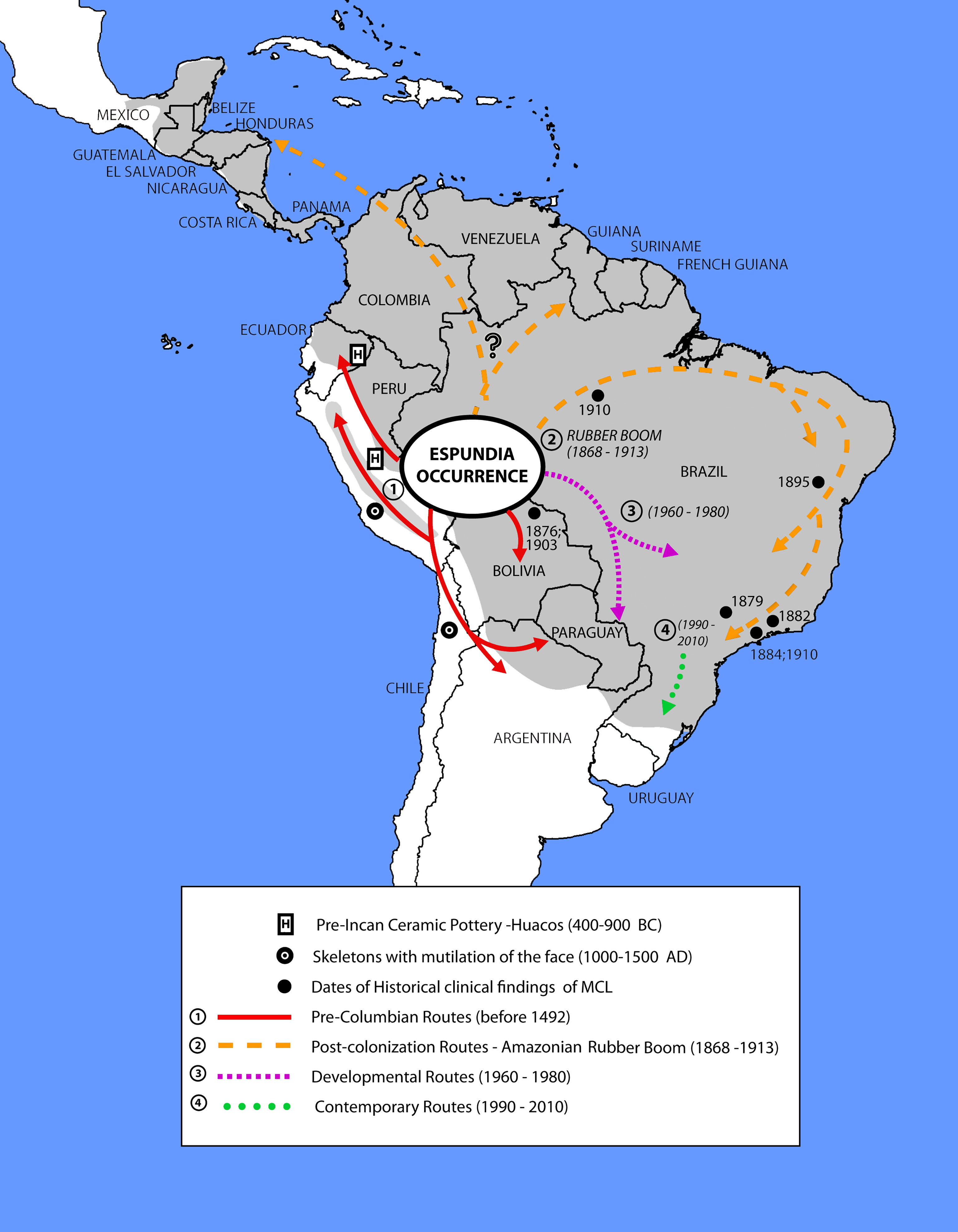
Figure 1 Archeological, paleopathological, and clinical/historical findings and possible dispersal routes for Leishmania (V.) braziliensis in the Americas. Adapted from (4, 8, 12, 63–66, 70).
Final Remarks
In endemic areas, the high proportion of individuals with non-symptomatic infection, even those considered treated and cured, but at risk of reactivation, in addition to the growing importance of domestic transmission, suggest convincing circumstantial evidence of the anthroponotic transmission of Leishmania (Viannia) (32).
Parasite persistence and the narrower genetic diversity of L. (V.) braziliensis isolates, found in humans (86–88) and with a wide geographic distribution (5), reinforce the evidence that the parasite was initially introduced and continues to be spread by the human host in environments that have been altered for a long time, as well as in recently deforested environments with high vector density.
This situation, in addition to sylvatic transmission, gives rise to new foci of the endemic, producing various transmission patterns: peri-sylvatic, rural, peri-urban and urban, aggravated by the peri-domiciliary presence of dogs, equids, and/or synanthropic animals (89). In both peri-domiciliary and domiciliary transmission, it is common for younger persons and women to be affected (8).
Thus far, samples from blood and skin fragments of R. rattus, captured in an endemic area in the state of Minas Gerais, were found positive for L. (V.) braziliensis in PCR-hybridization experiments (85). R. rattus, with eclectic habits, introduced into the Americas by European colonists, and synanthropic marsupials whose population grows with anthropic activities in the peri-domicile, could be a link between the sylvatic and peri-domiciliary environments. These animals could take the opposite route, carrying the Leishmania introduced by humans or dogs from the peri-domiciliary cycle to the sylvatic environment in a process similar to the introduction of the bubonic plague in the Americas, associated with R. rattus, establishing a secondary sylvatic cycle. In addition, L. (L.) infantum was introduced into the Americas during the colonization periods and nowadays such specie is found in domiciliary, peri-domiciliary, and sylvatic cycles infecting dogs, marsupials, rodents, and foxes, respectively (90).
In short, beginning with its Amazonian origin, the parasite’s dispersal across various biomes due to human migrations associated with high rates of subclinical infections, as well as the chronicity of the disease and parasite persistence in infected or treated humans, linked to the presence of sandflies and the high susceptibility of domestic dogs, are the facts that point to the anthropogenic spread of L. (V.) braziliensis in the Americas.
Data Availability Statement
The original contributions presented in the study are included in the article/supplementary material. Further inquiries can be directed to the corresponding author.
Author Contributions
MM conceived the idea of the study, designed the methodology and wrote the first draft of the manuscript. KM, AF, AOS, LM, and RP contributed to investigation, analyze, data interpretation and review of the manuscript. MM and AOS got research funding. All authors contributed to the article and approved the submitted version.
Funding
This study was funded by grants from Fundação Carlos Chagas de Amparo à Pesquisa do Estado do Rio de Janeiro (FAPERJ)/ CNE E-26/202.911/2015 (AdOS) and National Council for Scientific and Technological Development – CNPq 302.414/ 2018-5 (AdOS) and 308889/2017-7 (MCdAM).
Conflict of Interest
The authors declare that the research was conducted in the absence of any commercial or financial relationships that could be construed as a potential conflict of interest.
Publisher’s Note
All claims expressed in this article are solely those of the authors and do not necessarily represent those of their affiliated organizations, or those of the publisher, the editors and the reviewers. Any product that may be evaluated in this article, or claim that may be made by its manufacturer, is not guaranteed or endorsed by the publisher.
Acknowledgments
The authors wish to thank Dr. Hooman Momen for his suggestions and criticisms to the manuscript, to Saulo Feldman Marzochi for preparation and accessibility of the map (Figure 1), and Dr. Christopher Peterson for the English translation. The authors also thank the National Council of Scientific and Technological Development (CNPq) for Productivity Grants.
References
1. Davies CR, Reithinger R, Campbell-Lendrum D, Feliciangeli D, Borges R, Rodriguez N. The Epidemiology and Control of Leishmaniasis in Andean Countries. Cad Saude Publica (2000) 16:925–50. doi: 10.1590/s0102-311x2000000400013
2. World Health Organization. WHO | Global Leishmaniasis Update, 2006–2015: A Turning Point in Leishmaniasis Surveillance. Geneva: WHO (2017). Available at: http://www.who.int/leishmaniasis/resources/who_wer9238/en/.
3. Ministério da Saúde, Secretaria de Vigilância em Saúde. Manual De Vigilância Da Leishmaniose Tegumentar [Recurso Eletrônico]. Ministério da Saúde: Brasilia-DF (2017).
4. Bedoya-Pacheco SJ, Araujo-Melo MH, Valete-Rosalino CM, Pimentel MIF, Conceição-Silva F, Schubach AO, et al. Endemic Tegumentary Leishmaniasis in Brazil: Correlation Between Level of Endemicity and Number of Cases of Mucosal Disease. Am J Trop Med Hyg (2011) 84:901–5. doi: 10.4269/ajtmh.2011.10-0595
5. Cupolillo E, Brahim LR, Toaldo CB, de Oliveira-Neto MP, de Brito MEF, Falqueto A, et al. Genetic Polymorphism and Molecular Epidemiology of Leishmania (Viannia) Braziliensis From Different Hosts and Geographic Areas in Brazil. J Clin Microbiol (2003) 41:3126–32. doi: 10.1128/JCM.41.7.3126-3132.2003
6. Tojal da Silva AC, Cupolillo E, Volpini AC, Almeida R, Sierra Romero GA. Species Diversity Causing Human Cutaneous Leishmaniasis in Rio Branco, State of Acre, Brazil. Trop Med Int Health (2006) 11:1388–98. doi: 10.1111/j.1365-3156.2006.01695.x
7. Miranda L de FC, Pacheco R da S, Pimentel MIF, Salgueiro M de M, da Silva AF, de Mello CX, et al. Geospatial Analysis of Tegumentary Leishmaniasis in Rio De Janeiro State, Brazil From 2000 to 2015: Species Typing and Flow of Travelers and Migrants With Leishmaniasis. PloS Negl Trop Dis (2019) 13:e0007748. doi: 10.1371/journal.pntd.0007748
8. Marzochi MC de A, Marzochi KBF. Tegumentary and Visceral Leishmaniases in Brazil: Emerging Anthropozoonosis and Possibilities for Their Control. Cad Saúde Pública (1994) 10:S359–75. doi: 10.1590/S0102-311X1994000800014
9. Amato VS, Tuon FF, Siqueira AM, Nicodemo AC, Neto VA. Treatment of Mucosal Leishmaniasis in Latin America: Systematic Review. Am J Trop Med Hyg (2007) 77:266–74. doi: 10.4269/ajtmh.2007.77.266
10. Saravia NG, Segura I, Labrada LA, Weigle K, Giannini SH, Pacheco R, et al. Recurrent Lesions in Human Leishmania Braziliensis Infection—Reactivation or Reinfection? Lancet (1990) 336:398–402. doi: 10.1016/0140-6736(90)91945-7
11. Lainson R. The Neotropical Leishmania Species: A Brief Historical Review of Their Discovery, Ecology and Taxonomy. Rev Pan-Amaz Saúde (2010) 1:13–32. doi: 10.5123/S2176-62232010000200002
12. Altamirano-Enciso AJ, Marzochi MCA, Moreira JS, Schubach AO, Marzochi KBF. [on the Origin and Spread of Cutaneous and Mucosal Leishmaniasis, Based on Pre- and Post- Colombian Historical Source]. Hist Cienc Saude–Manguinhos (2003) 10:852–82. doi: 10.1590/S0104-59702003000300004
13. Nolder D, Roncal N, Davies CR, Llanos-Cuentas A, Miles MA. Multiple Hybrid Genotypes of Leishmania (Viannia) in a Focus of Mucocutaneous Leishmaniasis. Am J Trop Med Hyg (2007) 76:573–8. doi: 10.4269/ajtmh.2007.76.573
14. WHO Expert Committee on the Control of the Leishmaniases, World Health Organization eds. Control of the Leishmaniases: Report of a Meeting of the WHO Expert Committee on the Control of Leishmaniases. Geneva, Switzerland: World Health Organization (2010).
15. Brandão-Filho SP, Brito ME, Carvalho FG, Ishikaw EA, Cupolillo E, Floeter-Winter L, et al. Wild and Synanthropic Hosts of Leishmania (Viannia) Braziliensis in the Endemic Cutaneous Leishmaniasis Locality of Amaraji, Pernambuco State, Brazil. Trans R Soc Trop Med Hyg (2003) 97:291–6. doi: 10.1016/S0035-9203(03)90146-5
16. Maia-Elkhoury ANS, Magalhães Lima D, Salomón OD, Buzanovsky LP, Saboyá-Díaz MI, Valadas SYOB, et al. Interacción Entre Los Determinantes Medioambientales Y Socioeconómicos Para El Riesgo Para Leishmaniasis Cutánea En América Latina. Rev Panam Salud Pública (2021) 45:e49. doi: 10.26633/RPSP.2021.49
17. Gil JF, Nasser JR, Cajal SP, Juarez M, Acosta N, Cimino RO, et al. Urban Transmission of American Cutaneous Leishmaniasis in Argentina: Spatial Analysis Study. Am J Trop Med Hyg (2010) 82:433–40. doi: 10.4269/ajtmh.2010.09-0113
18. García AL, Parrado R, Rojas E, Delgado R, Dujardin J-C, Reithinger R. Leishmaniases in Bolivia: Comprehensive Review and Current Status. Am J Trop Med Hyg (2009) 80:704–11. doi: 10.4269/ajtmh.2009.80.704
19. Pirmez C, Marzochi MCA, Coutinho SG, Pirmez C, Marzochi MCA, Coutinho SG. Experimental Canine Mucocutaneous Leishmaniasis (Leishmania Braziliensis Braziliensis). Mem Inst Oswaldo Cruz (1988) 83:145–51. doi: 10.1590/S0074-02761988000200001
20. Reithinger R, Espinoza JC, Llanos-Cuentas A, Davies CR. Domestic Dog Ownership: A Risk Factor for Human Infection With Leishmania (Viannia) Species. Trans R Soc Trop Med Hyg (2003) 97:141–5. doi: 10.1016/S0035-9203(03)90101-5
21. Travi BL, Tabares CJ, Cadena H. Leishmania (Viannia) Braziliensis Infection in Two Colombian Dogs: A Note on Infectivity for Sand Flies and Response to Treatment. BioMed Rev Inst Nac Salud (2006) 26(Suppl 1):249–53. doi: 10.7705/biomedica.v26i1.1520
22. de Oliveira GM, Madeira M de F, Oliveira FS, Pacheco RS. PCR Associated With Molecular Hybridization Detects Leishmania (Viannia) Braziliensis in Healthy Skin in Canine Tegumentary Leishmaniasis. J Parasitol (2015) 101:91–4. doi: 10.1645/14-567.1
23. Ready PD. Sand Fly Evolution and its Relationship to Leishmania Transmission. Mem Inst Oswaldo Cruz (2000) 95:589–90. doi: 10.1590/S0074-02762000000400024
24. Gomes RF, Macedo AM, Pena SD, Melo MN. Leishmania (Viannia) Braziliensis: Genetic Relationships Between Strains Isolated From Different Areas of Brazil as Revealed by DNA Fingerprinting and RAPD. Exp Parasitol (1995) 80:681–7. doi: 10.1006/expr.1995.1084
25. Lopes UG, Momen H, Grimaldi G, Marzochi MC, Pacheco RS, Morel CM. Schizodeme and Zymodeme Characterization of Leishmania in the Investigation of Foci of Visceral and Cutaneous Leishmaniasis. J Parasitol (1984) 70:89–98. doi: 10.2307/3281930
26. Rougeron V, De Meeus T, Hide M, Waleckx E, Bermudez H, Arevalo J, et al. Extreme Inbreeding in Leishmania Braziliensis. Proc Natl Acad Sci (2009) 106:10224–9. doi: 10.1073/pnas.0904420106
27. Ishikawa E a Y, Silveira FT, Magalhães ALP, Guerra júnior RB, Melo MN, Gomes R, et al. Genetic Variation in Populations of Leishmania Species in Brazil. Trans R Soc Trop Med Hyg (2002) 96(Suppl 1):S111–21. doi: 10.1016/s0035-9203(02)90061-1
28. Lima ACVM da R. Estudo Da Variabilidade Genética De Leishmania (Viannia) Braziliensis Vianna, 1911 De Diferentes Regiões do Brasil (2010). Available at: http://www.bibliotecadigital.ufmg.br/dspace/handle/1843/SAGF-8HCK3K (Accessed December 1, 2020).
29. Boité MC, Mauricio IL, Miles MA, Cupolillo E. New Insights on Taxonomy, Phylogeny and Population Genetics of Leishmania (Viannia) Parasites Based on Multilocus Sequence Analysis. PloS Negl Trop Dis (2012) 6:e1888. doi: 10.1371/journal.pntd.0001888
30. Motoie G, Ferreira GEM, Cupolillo E, Canavez F, Pereira-Chioccola VL. Spatial Distribution and Population Genetics of Leishmania Infantum Genotypes in São Paulo State, Brazil, Employing Multilocus Microsatellite Typing Directly in Dog Infected Tissues. Infect Genet Evol (2013) 18:48–59. doi: 10.1016/j.meegid.2013.04.031
31. Saberi R, Fakhar M, Mohebali M, Anvari D, Gholami S. Global Status of Synchronizing Leishmania RNA Virus in Leishmania Parasites: A Systematic Review With Meta-Analysis. Transbound Emerg Dis (2019) 66:2244–51. doi: 10.1111/tbed.13316
32. Martínez-Valencia AJ, Daza-Rivera CF, Rosales-Chilama M, Cossio A, Casadiego Rincón EJ, Desai MM, et al. Clinical and Parasitological Factors in Parasite Persistence After Treatment and Clinical Cure of Cutaneous Leishmaniasis. PloS Negl Trop Dis (2017) 11:e0005713. doi: 10.1371/journal.pntd.0005713
33. Mendonça MG, de Brito MEF, Rodrigues EHG, Bandeira V, Jardim ML, Abath FGC. Persistence of Leishmania Parasites in Scars After Clinical Cure of American Cutaneous Leishmaniasis: Is There a Sterile Cure? J Infect Dis (2004) 189:1018–23. doi: 10.1086/382135
34. Schubach A, Marzochi MC, Cuzzi-Maya T, Oliveira AV, Araujo ML, Oliveira AL, et al. Cutaneous Scars in American Tegumentary Leishmaniasis Patients: A Site of Leishmania (Viannia) Braziliensis Persistence and Viability Eleven Years After Antimonial Therapy and Clinical Cure. Am J Trop Med Hyg (1998) 58:824–7. doi: 10.4269/ajtmh.1998.58.824
35. Schubach A, Haddad F, Neto MP-O, Degrave W, Pirmez C, Grimaldi G Jr, et al. Detection of Leishmania DNA by Polymerase Chain Reaction in Scars of Treated Human Patients. J Infect Dis (1998) 178:911–4. doi: 10.1086/515355
36. Vergel C, Palacios R, Cadena H, Posso CJ, Valderrama L, Perez M, et al. Evidence for Leishmania (Viannia) Parasites in the Skin and Blood of Patients Before and After Treatment. J Infect Dis (2006) 194:503–11. doi: 10.1086/505583
37. Camera P de O, Junger J, Pires F do ESS, Mattos M, Oliveira-Neto MP, Fernandes O, et al. Haematogenous Dissemination of Leishmania (Viannia) Braziliensis in Human American Tegumentary Leishmaniasis. Trans R Soc Trop Med Hyg (2006) 100:1112–7. doi: 10.1016/j.trstmh.2006.02.014
38. Coutinho SG, Pirmez C, Da-Cruz AM. Parasitological and Immunological Follow-Up of American Tegumentary Leishmaniasis Patients. Trans R Soc Trop Med Hyg (2002) 96:S173–8. doi: 10.1016/S0035-9203(02)90072-6
39. Guevara P, Rojas E, Gonzalez N, Scorza JV, Añez N, Valera M, et al. Presence of Leishmania Braziliensis in Blood Samples From Cured Patients or at Different Stages of Immunotherapy(1994) (Accessed January 8, 2021).
40. Quintella LP, Cuzzi T, Madeira M de F, Okamoto T, Schubach A de O. Immunoperoxidase Technique Using an Anti-Leishmania (L.) Chagasi Hyperimmune Serum in the Diagnosis of Culture-Confirmed American Tegumentary Leishmaniasis. Rev Inst Med Trop São Paulo (2009) 51:83–6. doi: 10.1590/S0036-46652009000200005
41. Fagundes A, Marzochi MCA, Perez M, Schubach A, Ferreira A, Silva JP, et al. Skin Reactivity to Thimerosal and Phenol-Preserved Montenegro Antigen in Brazil. Acta Trop (2007) 101:25–30. doi: 10.1016/j.actatropica.2006.11.007
42. Ivonise F, Cibele A, Olívia B, Clarissa BA, Lucas PC, Roque PA, et al. Epidemiologic and Immunologic Findings for the Subclinical Form of Leishmania Braziliensis Infection. Clin Infect Dis (2002) 34:e54–8. doi: 10.1086/340261
43. Silveira FT, Lainson R, Shaw JJ, Ishikawa EA, Souza AA, Braga RR. Sensitivity of the Culture of Circulating Leukocytes in the Detection of Leishmania in the Peripheral Blood of Patients With Tegumentary Leishmaniasis. Rev Soc Bras Med Trop (1989) 22:143–6. doi: 10.1590/s0037-86821989000300006
44. Biagi F. [Some Comments on Leishmaniasis and its Agents: Leishmania Tropica Mexicana, New Subspecies]. Medicina (Mex) (1953) 33:401–6.
45. Floch H. Leishmania Tropica Guyanensis N.Sp., Pathogenic Agent of Guyanese and Central American Cutaneous Laishmaniasis. Bull Soc Pathol Exot Filiales (1954) 47:784–7.
46. Forattini OP. Sobre Os Reservatórios Naturais De Leishmaniose Tegumentar Americana(1960) (Accessed December 15, 2020).
47. Forattini OP, Pattoli DB, Rabello EX, Ferreira OA. [Natural Infections of Wild Mammals in an Endemic Area of Tegmental Leishmaniasis in the State of São Paulo, Brazil]. Rev Saude Publica (1972) 6:255–61. doi: 10.1590/S0034-89101972000300003
48. Forattini OP, Pattoli DB, Rabello EX, Ferreira OA. [Notes on Natural Infection of Oryzomys Capito Laticeps in an Euzootic Focus of Cutaneous Leishmaniasis in the Estado De São Paulo, Brazil]. Rev Saude Publica (1973) 7:181–4. doi: 10.1590/s0034-89101973000200010
49. Montoya-Lerma J, Palacios R, Osorio L, Jaramillo C, Cadena H. Further Evidence of Humans as Source of Leishmania Viannia for Sandflies. Mem Inst Oswaldo Cruz (1998) 93:735–6. doi: 10.1590/S0074-02761998000600006
50. Rojas E, Scorja JV. Xenodiagnostico Con Lutzomyia Youngi En Casos Venezolanos De Leishmaniasis Cutaned Por Leishmania Braziliensis. Mem Inst Oswaldo Cruz (1989) 84:29–34. doi: 10.1590/S0074-02761989000100006
51. Shaw J. The Leishmaniases–Survival and Expansion in a Changing World. A Mini-Review. Mem Inst Oswaldo Cruz (2007) 102:541–7. doi: 10.1590/S0074-02762007000500001
52. Oliveira CCG, Lacerda HG, Martins DRM, Barbosa JDA, Monteiro GR, Queiroz JW, et al. Changing Epidemiology of American Cutaneous Leishmaniasis (ACL) in Brazil: A Disease of the Urban–Rural Interface. Acta Trop (2004) 90:155–62. doi: 10.1016/j.actatropica.2003.11.011
53. Ampuero J, Urdaneta M, Macêdo V de O. Risk Factors for Cutaneous Leishmaniasis Transmission in Children Aged 0 to 5 Years in an Endemic Area of Leishmania (Viannia) Braziliensis. Cad Saúde Pública (2005) 21:161–70. doi: 10.1590/S0102-311X2005000100018
54. Castellucci L, Cheng LH, Araújo C, Guimarães LH, Lessa H, Machado P, et al. Familial Aggregation of Mucosal Leishmaniasis in Northeast Brazil. Am J Trop Med Hyg (2005) 73:69–73. doi: 10.4269/ajtmh.2005.73.69
55. Falqueto A, Coura JR, Barros GC, Grimaldi Filho G, Sessa PA, Carias VR, et al. [Participation of the Dog in the Cycle of Transmission of Cutaneous Leishmaniasis in the Municipality of Viana, State of Espirito Santo, Brazil]. Mem Inst Oswaldo Cruz (1986) 81:155–63. doi: 10.1590/s0074-02761986000200004
56. de Oliveira AC, Figueiredo FB, Silva VL, Santos FN, de Souza MB, de Madeira MF, et al. Canine Visceral Leishmaniasis Case Investigation in the Jacare Region of Niteroi, Rio De Janeiro, Brazil. Rev Inst Med Trop São Paulo (2015) 57:325–32. doi: 10.1590/S0036-46652015000400009
57. Santos EG, Marzochi MC, Conceição NF, Brito CM, Pacheco RS. Epidemiological Survey on Canine Population With the Use of Immunoleish Skin Test in Endemic Areas of Human American Cutaneous Leishmaniasis in the State of Rio De Janeiro, Brazil. Rev Inst Med Trop Sao Paulo (1998) 40:41–7. doi: 10.1590/S0036-46651998000100009
58. dos Santos GPL, Sanavria A, Marzochi MC de A, dos Santos EGOB, Silva VL, da Pacheco RS, et al. Prevalência Da Infecção Canina Em Áreas Endêmicas De Leishmaniose Tegumentar Americana, do Município De Paracambi, Estado do Rio De Janeiro, No Período Entre 1992 E 1993. Rev Soc Bras Med Trop (2005) 38:161–6. doi: 10.1590/S0037-86822005000200007
59. Aguilar CM, Rangel EF, Garcia L, Fernandez E, Momen H, Grimaldi Filho G, et al. Zoonotic Cutaneous Leishmaniasis Due to Leishmania (Viannia) Braziliensis Associated With Domestic Animals in Venezuela and Brazil. Mem Inst Oswaldo Cruz (1989) 84:19–28. doi: 10.1590/s0074-02761989000100005
60. Barbosa-Santos EG, Marzochi MC, Urtado W, Queirós F, Chicarino J, Pacheco RS. Leishmaniasis Disseminated by Leishmania Braziliensis in a Mare (Equus Cabalus) Immunotherapy and Chemotherapy Assays. Mem Inst Oswaldo Cruz (1994) 89:217–20. doi: 10.1590/s0074-02761994000200018
61. Schubach TMP, Figueiredo FB, Pereira SA, Madeira MF, Santos IB, Andrade MV, et al. American Cutaneous Leishmaniasis in Two Cats From Rio De Janeiro, Brazil: First Report of Natural Infection With Leishmania (Viannia) Braziliensis. Trans R Soc Trop Med Hyg (2004) 98:165–7. doi: 10.1016/S0035-9203(03)00040-3
62. Yoshida EL, Correa FM, Marques SA, Stolf HO, Dillon NL, Momen H, et al. Human, Canine and Equine (Equus Caballus) Leishmaniasis Due to Leishmania Braziliensis (= L. Braziliensis Braziliensis) in the South-West Region of São Paulo State, Brazil. Mem Inst Oswaldo Cruz (1990) 85:133–4. doi: 10.1590/s0074-02761990000100026
64. Escomel E. La Leishmaniose Américaine Etles Leishmanioses En Amériques. Bull Soc Path Exot (1929) 22:35–46.
65. Altamirano-Enciso AJ, Moreira JS, Marzochi MCA. Lesión Lítica Craniana Por Leishmaniasis En Makat-Tampu Durante El Imperio Inca: Silos XV – XVI, Valle Del Bajo Rímac, Peru. Rev Museu Arq Etnol (2001) 11:227–42. doi: 10.11606/issn.2448-1750.revmae.2001.109420
66. Costa MA, Matheson C, Iachetta L, Llagostera A, Appenzeller O. Ancient Leishmaniasis in a Highland Desert of Northern Chile. PloS One (2009) 4:e6983. doi: 10.1371/journal.pone.0006983
67. Marsteller SJ, Torres-Rouff C, Knudson KJ. Pre-Columbian Andean Sickness Ideology and the Social Experience of Leishmaniasis: A Contextualized Analysis of Bioarchaeological and Paleopathological Data From San Pedro De Atacama, Chile. Int J Paleopathol (2011) 1:24–34. doi: 10.1016/j.ijpp.2011.02.001
69. Momen H, Cupolillo E. Speculations on the Origin and Evolution of the Genus Leishmania. Mem Inst Oswaldo Cruz (2000) 95:583–8. doi: 10.1590/S0074-02762000000400023
70. Rabello E. Contribuições Ao Estudo Da Leishmaniose Tegumentar No Brasil. I. Histórico E Sinonímia. Ann Bras Dermatol Syphilog (1925) 1:3–31.
71. Thomaz-Soccol V, Lanotte G, Rioux JA, Pratlong F, Martini-Dumas A, Serres E. Monophyletic Origin of the Genus Leishmania Ross, 1903. Ann Parasitol Hum Comp (1993) 68:107–8.
72. do Vale ECS, Furtado T. Tegumentary Leishmaniasis in Brazil: A Historical Review Related to the Origin, Expansion and Etiology. Bras Dermatol (2005) 80:421–8. doi: 10.1590/S0365-05962005000400015
73. Weinstein B. The Amazon Rubber Boom, 1850-1920. Stanford - CA: Stanford University Press (1983).
74. Sousa AQ, Pearson R. Drought, Smallpox, and Emergence of Leishmania Braziliensis in Northeastern Brazil. Emerg Infect Dis (2009) 15:916–21. doi: 10.3201/eid1506.071331
75. Dean W. A Luta Pela Borracha No Brasil: Um Estudo De História Ecológica. 1a. São Paulo: Nobel (1989).
76. González G, Arce Queirolo A. Leishmaniosis.I. Historia De La Leishmaniosis En El Paraguay. Rev Médica del Paraguay (1955) 1:65–8.
77. Marzochi MC de A, Coelho R de B, Soares DA, Zeitune JMR, Muarrek FJ, Cecchini R, et al. Hepatic Carcinogenesis in the Northern Part of Parana State and the Indiscriminate Use of Pesticides. I. Introduction to a Research Program (1976) (Accessed December 1, 2020).
78. Marzochi MC, Coutinho SG, Sabroza PC, de Souza WJ. Indirect Immunofluorescence Reaction and Intradermoreaction for American Cutaneous Leishmaniasis in Residents of the Jacarepagua Region (Rio De Janeiro). Comparative Study of Results Observed in 1974 and 1978. Rev Inst Med Trop Sao Paulo (1980) 22:149–55.
79. Passos VMA, Falcão AL, Marzochi MCA, Gontijo CMF, Dias ES, Barbosa-Santos EGO, et al. Epidemiological Aspects of American Cutaneous Leishmaniasis in a Periurban Area of the Metropolitan Region of Belo Horizonte, Minas Gerais, Brazil. Mem Inst Oswaldo Cruz (1993) 88:103–10. doi: 10.1590/S0074-02761993000100016
80. Sabroza PC. O Domicílio Como Fator De Risco Na Leishmaniose Tegumentar Americana. Município do Rio de Janeiro: Estudo Epidemiológico em Jacarepaguá (1983).
81. Chaves LF, Cohen JM, Pascual M, Wilson ML. Social Exclusion Modifies Climate and Deforestation Impacts on a Vector-Borne Disease. PloS Negl Trop Dis (2008) 2:e176. doi: 10.1371/journal.pntd.0000176
82. Pacheco RS, Barbosa-Santos EGO, Brito CMM, Pires MQ, Marzochi MCA. Epidemiological and Genotypical Mapping of Human Leishmania (Viannia) Braziliensis in Paraguay. J Protozool Res (1999) 9:76–87. doi: 10.32268/jprotozoolres.9.3_76
83. Vora N. Impact of Anthropogenic Environmental Alterations on Vector-Borne Diseases (2008) (Accessed January 18, 2021).
84. Da-Cruz AM, Machado ES, Menezes JA, Rutowitsch MS, Coutinho SG. Cellular and Humoral Immune Responses of a Patient With American Cutaneous Leishmaniasis and AIDS. Trans R Soc Trop Med Hyg (1992) 86:511–2. doi: 10.1016/0035-9203(92)90089-U
85. Oliveira FS, Pirmez C, Pires MQ, Brazil RP, Pacheco RS. PCR-Based Diagnosis for Detection of Leishmania in Skin and Blood of Rodents From an Endemic Area of Cutaneous and Visceral Leishmaniasis in Brazil. Vet Parasitol (2005) 129:219–27. doi: 10.1016/j.vetpar.2005.01.005
86. Baptista C, Schubach AO, Madeira MF, Leal CA, Pires MQ, Oliveira FS, et al. Leishmania (Viannia) Braziliensis Genotypes Identified in Lesions of Patients With Atypical or Typical Manifestations of Tegumentary Leishmaniasis: Evaluation by Two Molecular Markers. Exp Parasitol (2009) 121:317–22. doi: 10.1016/j.exppara.2008.12.006
87. de Oliveira FS, Valete-Rosalino CM, de Oliveira Schubach A, de Fátima Madeira M, da Silva Pacheco R. Genetic Polymorphism in Leishmania (Viannia) Braziliensis Detected in Mucosal Leishmaniasis of HIV-Infected and non-HIV-Infected Patients. Trans R Soc Trop Med Hyg (2012) 106:683–7. doi: 10.1016/j.trstmh.2012.07.007
88. de Oliveira GM, Madeira M de F, Oliveira FS, Pires MQ, Pacheco R da S. Canine Cutaneous Leishmaniasis: Dissemination and Tissue Tropism of Genetically Distinct Leishmania (Viannia) Braziliensis Populations. Vet Med Int (2013) 2013:1–5. doi: 10.1155/2013/982183
89. Marzochi MCA, Marzochi KBF. Leishmanioses Em Áreas Urbanas. Rev Soc Bras Med Trop (1997) 30:162–4.
Keywords: Leishmania (Viannia) braziliensis, mucocutaneous leishmaniasis, parasite persistence, human migrations, dogs, expansion in the Americas, anthroponosis, control
Citation: Marzochi MCdA, Marzochi KBF, Fagundes A, Schubach AdO, Miranda LdFC and Pacheco RdS (2021) Anthropogenic Dispersal of Leishmania (Viannia) braziliensis in the Americas: A Plausible Hypothesis. Front. Trop. Dis 2:723017. doi: 10.3389/fitd.2021.723017
Received: 09 June 2021; Accepted: 24 August 2021;
Published: 14 September 2021.
Edited by:
Angela H. Lopes, Federal University of Rio de Janeiro, BrazilReviewed by:
Ricardo Fujiwara, Federal University of Minas Gerais, BrazilSeray Toz, Ege University Medical Faculty Department of Parasitology, Turkey
Robert McMaster, University of British Columbia, Canada
Copyright © 2021 Marzochi, Marzochi, Fagundes, Schubach, Miranda and Pacheco. This is an open-access article distributed under the terms of the Creative Commons Attribution License (CC BY). The use, distribution or reproduction in other forums is permitted, provided the original author(s) and the copyright owner(s) are credited and that the original publication in this journal is cited, in accordance with accepted academic practice. No use, distribution or reproduction is permitted which does not comply with these terms.
*Correspondence: Mauro Célio de Almeida Marzochi, mauromarzochi@uol.com.br