High Ankle Injuries or Syndesmosis injuries are important to be diagnosed correctly as the management and return from injury is very different to that of the common lateral ligament ankle sprain.
Anatomy
The lateral ligaments of an ankle and the syndesmosis ligament (anteriorly) are in close anatomical proximity to each other. This is why a lateral ligament ankle sprain can be mistaken for a syndesmosis injury.
Syndesmosis complex (in RED) consists of the anterior inferior tibiofibular ligament (AITFL), posterior inferior tibiofibular ligament (PITFL), interosseous ligament (IOL), and the inferior transverse ligament (ITL). Its function is to hold the lower leg bones together – the tibia and fibula, so the ankle joint remains stable.
The lateral ligaments (in BLUE) consist of the anterior talofibular ligament (ATFL), calcaneofibular ligament (CFL) and posterior talofibular ligament (PTFL).

How Are They Injured?
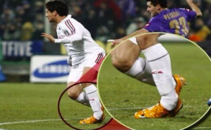
Syndesmosis Injury – Forced External Rotation of the ankle. Most commonly the force from an opponent landing on the outside of the lower leg, causing the ankle to be forced into external rotation (toes pointed outwards). This causes the fibula and tibia to be separated at the ankle and injuring the syndesmosis complex.


Lateral Ligament Injury – Ankle Inversion +/- Plantar Flexion. Most commonly from a landing from a jump or changing direction causing the ankle to roll inwards. Some other factors can contribute to this cause are landing from a jump onto an opponent’s foot and rolling off it or rolling an ankle because of a divot in the ground. This causes the lateral ligaments to stretch and be injured.
Diagnosis of the Injuries
Important and accurate diagnosis is required by a trained Sports & Exercise Physiotherapist as the management varies between syndesmosis injuries versus lateral ligament injuries and the severity of if the injuries.
These injuries are diagnosed by understanding how the patient injured themselves (See pictures above). A clinical examination is performed by a trained physiotherapist, who will differentiate the two types of injuries and the severity of the injuries.
Syndesmosis injuries are classified into 4 grades:
Grade 1: Isolated injury to AITFL
Grade 2: Injury to AITFL and IOL
Grade 3: Injury to AITFL, IL and PITFL
Grade 4: Injury to AITFL, IL, PITFL and Deltoid Ligament.
Correct diagnosis can and should be confirmed with a MRI if suspicious of more than a grade 2 injury. Of the radiographic tools, MRI has been found to have the highest specificity and sensitivity – meaning the most accurate form of imaging available. X-Ray is used to rule out fractures/breaks in bones. The reliability of x-ray, particularly using stress radiographs to diagnose injury to the ankle syndesmosis is controversial.
Lateral ligament most commonly injured is the ATFL and then the CFL. PTFL are less likely to sustain damaging loads. Combination of the ligaments can also be injured.
Lateral Ligament Injuries are classified into 3 grades:
Grade 1: Microscopic injury without a partial tear
Grade 2: Macroscopic injury - Partial tear to ligament
Grade 3: Complete rupture to the ligament
These all present with different varying grades of symptoms. X-Ray can be used the exclude any acute bony injury and MRI can confirm the ligaments injured and the grading of these ligaents. MRI is usually only performed in a more serious presentations or where suspicion to other structures of the ankle such as talar dome may be injured.
Management
Syndesmosis Injuries
Syndesmosis injuries require a period of biological healing for the ligament before loading occurs. This ligament is loaded through weight-bearing and walking and therefore requires a period of time of off-load to allow healing to occur.
Usually grade 1 and grade 2 injuries are managed without surgery. They are put in a boot and are non-weight-bearing for a period of time depending on the grade. Management of swelling is important with compression, massage and ice. They are then weaned out of the boot and graded ROM, strength, balance of the ankle/foot is performed. This progresses to more functional rehab such as plyometrics, running mechanics, running and sport/exercise specific rehab when able. The rehab timelines for a grade 1-2 is usually 5-10 weeks depending on the patient’s presentation.
Grade 3 injuries and sometimes grade 2 injuries can be surgically stabilised by a tight rope or screws. Rehab management again is a graded process working on ROM, strength, balance and functional progressions. The rehab timeline for a tightrope surgical stabilisation is usually 8-16 weeks.
Lateral Ligament Injuries
Management is similar to that of a syndesmosis ankle injury however rehab timelines are far quicker because the ligaments are non-weight bearing ligaments. Rehab timelines are purely based on competency-based progressions where, if the patient can walk with minimal symptoms, get them to walk. Get them doing as much ROM, strength and functional rehab within what symptoms allow.
Rehab timelines are dependent on how many of the lateral ligaments are injured at once. If ATFL ligament injured in isolation the time frames may be from 3 days to 6 weeks depending on the severity/grade of injury. With two ligaments injured usually ATFL and CFL, usually this is a more severe injury may be 2-8 weeks.
What do you do if you have an ankle injury?
Book in for a consultation with one of our physiotherapists, so a correct diagnosis can be made to ensure a safe and timely return to your activity or sport.